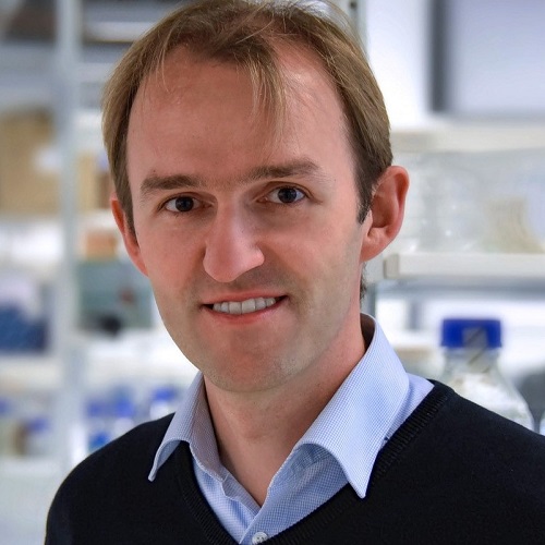
Dominik Paquet

iPSC- and CRISPR-based brain tissue models recapitulate key features of neurodegenerative and neurovascular diseases
Background
Brain research heavily depends on models recapitulating key aspects of human brain physiology and disease pathology. Human iPSCs have great potential to complement existing rodent disease models, as they allow directly studying affected human cell types. In addition, recent developments in CRISPR genome editing revolutionized how impacts of genetic alterations on disease formation can be investigated. Co-culture of disease-relevant iPSC-derived cells with disease-relevant mutations enables studying complex phenotypes involving cellular crosstalk.
Methods
By combining iPSC‑, CRISPR- and tissue engineering technologies, we established new brain tissue models for AD and FTD using iPSC-derived cortical neurons, astrocytes, microglia, and oligodendrocytes, as well as a microfluidic model of the neurovascular unit (NVU) based on co-culture of endothelial cells, mural cells and astrocytes
Results
Our technology provides highly controllable and reproducible 3‑dimensional tissues with typical cell morphologies and functional features, including widespread synapse formation, spontaneous and induced electrical activity, network formation, microglial ramification, tiling and phagocytosis, as well as formation of barrier-containing vessels interacting with astrocytic end feet for the NVU model. Interaction between neurons, glia and vascular cells become evident on morphological and functional levels. The models can be long-term cultured in a postmitotic state without proliferation or cell death, thus providing a more controllable, reproducible, and long-lived alternative to cortical organoids currently used for 3D disease modelling.
CRISPR-engineering of disease-causing mutations for AD, FTD, or a neurovascular disease induced characteristic late-stage phenotypes, including protein misfolding and aggregation for AD/FTD models, or barrier impairment for models of the NVU.
Conclusions
We expect that our models will enable studies elucidating novel, potentially human-specific pathomechanisms and provide a human framework for translation and screening.
Biography
Dr. Dominik Paquet received his PhD in 2009 from the Ludwig-Maximilians-University (LMU) in Munich, Germany, where he successfully developed the first transgenic zebrafish model of Tauopathies with Christian Haass and demonstrated its suitability for in vivo drug screening and real-time in vivo imaging of disease processes. During his postdoctoral work with Marc Tessier-Lavigne at The Rockefeller University in New York City, which was supported by the German Academy of Sciences Leopoldina and a New York Stem Cell Foundation Druckenmiller Fellowship, Dr. Paquet built up a new induced pluripotent stem cell (iPSC) laboratory at Rockefeller and developed efficient technologies to edit the genome of iPSCs by CRISPR/Cas9 editing to study Alzheimer’s and other neurodegenerative diseases in human brain cells. Dr. Paquet established the PaquetLab (www.isd-research.de/PaquetLab) at the Institute for Stroke and Dementia Research (ISD) of LMU Munich in 2017 and currently serves as Professor of Neurobiology and core member of Synergy (www.synergy-munich.de), a leading Research Cluster of the Excellence Initiative of the German research funding organization. Using his broad background in neurodegenerative disease research, with specific training and expertise in molecular, stem cell and neurobiology, he leads an interdisciplinary team of neurobiologists, biochemists, and stem cell biologists. The PaquetLab uses cutting-edge technologies such as CRISPR genome editing, iPSC differentiation and human 3D tissue engineering to develop a new generation of human brain tissue models to elucidate molecular functions of the human brain and mechanisms leading to neurodegenerative and neurovascular diseases.
